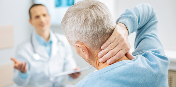
Degenerative cervical pathology is a common condition that affects the spine in the neck area, causing pain, stiffness and various neurological symptoms that can limit the quality of life of people who suffer from it.
One of the options to repair the condition is anterior cervical arthrodesis, a surgical procedure that consists of fusing two vertebrae, preventing movement and making the pain disappear by acting on the anterior part of the vertebra. Various techniques can be used for this.
Traditional anterior cervical arthrodesis techniques restored the function of the removed cervical disc using three elements: the patient’s own bone, titanium or PEEK (thermoplastic) cages filled with materials that promoted fusion, and cervical plates to facilitate the integration of the prostheses.
However, the appearance of screw cages is a remarkable milestone in the innovation of anterior cervical arthrodesis.
The main difference lies in the minimal invasion that characterises this technique, which translates into lower risks, shorter recovery times and more satisfactory results.
Compared to any disc substitute (non-screw cages or bone grafts), screw cages allow greater contact between the fused vertebrae in addition to added structural support that maintains the height of the disc space, facilitating bone integration.
Compared to classic cervical plates (often accompanied by non-screw cages or bone grafts), the main advantage of screw cages is a zero sagittal profile, that is, by being integrated into the vertebrae, they don’t occupy prevertebral space, avoiding the formation of osteophytes (bony protuberances in the joints) and the appearance of mechanical dysphagia (difficulty in swallowing when compressing the oesophagus). Plus, screw cages require less space for implantation, resulting in a much smaller wound and less need for traction/separation of neighbouring structures (oesophagus).
In summary, cervical screw cages represent an advanced and effective therapeutic option in the treatment of degenerative cervical pathology, providing patients with stability, correct bone integration, prevention of complications and a faster and less painful recovery. This minimally invasive technique has revolutionized the field of spinal surgery, being a safe and much more effective alternative to achieve the proposed objectives (recovering the curvature of the spine in the cervical region) with a modern and less invasive approach.
This technique, performed by an experienced surgical team, considerably reduces the risks associated with surgery, favours a prompt recovery by minimising the impact on the surrounding tissues, decreases blood loss and facilitates a shorter hospital stay and faster recovery compared to conventional techniques.
Dr. José Vicente Mollá
Neurosurgeon specialising in spinal surgery
Vithas Alicante Perpetuo Socorro Hospital